Research Equipment
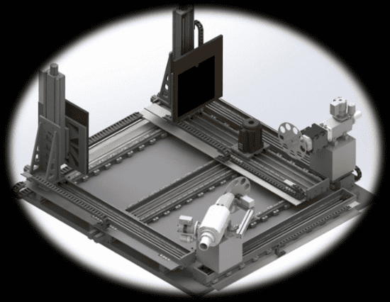
Advanced workstation computer X-ray tomography combines two pairs of " X-ray tube - Display " (the Dual Source CT - DSCT ) in an orthogonal arrangement , which has a two-fold acceleration of the process of collecting data for tomographic reconstruction . The workplace has a fully motorized axes for distance setting "RTG - sample - detector " . This makes it possible to change the magnification of about 1.2 times to 100 times . Given the size of the detector pixels can change the resolution CT reconstruction from 0.2 millimeters to micrometers , the size of the detector. Very stable high resolution is possible also with regard to the use of anti-vibration table, on which the whole assembly is placed , and last but not least, thanks to the installation of high precision rotary table tomography.
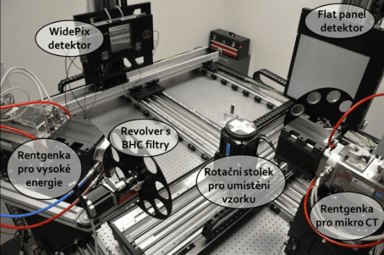
Another advantage is the ability to work DSCT parallel imaging an object in two spectra of X-ray radiation ( so-called dual energy radiography ) . This procedure allows you to highlight differences between the material components to the full X-ray spectrum similar to the attenuation of the radiation . If the sample consists of only two materials , these materials can be clearly distinguished. For multicomponent materials can only emphasize the differences.
Departments will also be equipped with detectors type Medipix / Timepix that allow you to set the display range in 4 -bit range. This will be the combination of two successive detectors collecting data 2x16 to get energy channels and tomographic reconstructions clearly distinguish up to 32 different materials. This work is in DSCT completely unique , even in the world
| Model | Minimum spot size | Maximum power on the target | Maximum energy | Information |
|---|---|---|---|---|
| XWT 160 TCHR | 1 µm | 3 W | 160 keV | For micro- CT measurements |
| XWT 240 SE | 4 µm | 280 W | 240 keV | For high power |
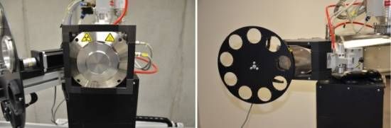
X-ray tube XWT 240 SE is designed for high- CT measurement and is therefore suitable for large samples or samples that strongly absorb X-rays . The diameter of the radiation beam based on the X-ray tube , the spot size is 4 µm. X-ray tube produces radiation with a maximum energy of 240 keV and its maximum power is 280 W. X-ray tube XWT 160 TCHR is designed for micro- CT measurement and is therefore more suitable for smaller samples and those that do not absorb X-rays too strongly , because it produces less radiation of maximum energy and 160 keV. This X-ray tube operates in two modes depending on the desired application . High resolution mode provides a spot size of 1 µm , but the maximum power on the target of only 3 watts High energy mode provides a spot size of 4 µm , but the maximum power is 10 W.
| Model | Size | Matrix of pixels | Size of pixels | Information |
|---|---|---|---|---|
| Flat panel | 400x400 mm | 2048x2048 | 200x200 µm | Indirect signal conversion |
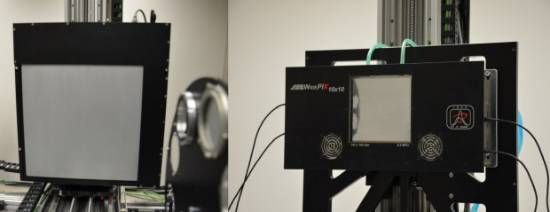
| WidePix | 140x140 mm | 2560x2560 | 55x55 µm | Unique in the world |
| Model | Resolution | Maximum speed | Axial Weight Capacity | Radial Weight Capacity |
|---|---|---|---|---|
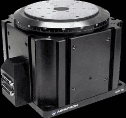
| ABRT-150-AS | 0.55 arcseconds | 1200 rpm | 20 kg | 3 kg |
| APR-150-DR-135 | 0.08 arcseconds | 600 rpm | 45 kg | 32 kg |